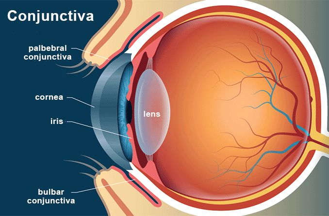
Conjunctiva of the eye

An important structure on the surface of the eye is one you can’t easily see — it’s called the conjunctiva.
Conjunctiva definition
The conjunctiva is the clear, thin membrane that covers part of the front surface of the eye and the inner surface of the eyelids. It has two segments:
Bulbar conjunctiva. This portion of the conjunctiva covers the anterior part of the sclera (the “white” of the eye). The bulbar conjunctiva stops at the junction between the sclera and cornea; it does not cover the cornea.
Palpebral conjunctiva. This portion covers the inner surface of both the upper and lower eyelids. (Another term for the palpebral conjunctiva is tarsal conjunctiva.)
The bulbar and palpebral conjunctiva are continuous (see illustration). This feature makes it impossible for a contact lens (or anything else) to get lost behind your eye.
SEE RELATED: Contact lenses and pink eye
Conjunctiva function
The primary functions of the conjunctiva are:
Keep the front surface of the eye moist and lubricated.
Keep the inner surface of the eyelids moist and lubricated so they open and close easily without friction or causing eye irritation.
Protect the eye from dust, debris and infection-causing microorganisms.
The conjunctiva has many small blood vessels that provide nutrients to the eye and lids. It also contains special cells that secrete a component of the tear film to help prevent dry eye syndrome.
Conjunctiva problems
A number of conditions can affect the conjunctiva. Among the more common conjunctival problems are:
Conjunctivitis. Also called pink eye, this is inflammation of the conjunctiva. It can have several causes.
Conjunctival pallor. This is an unhealthy pale appearance to the palpebral conjunctiva that can be a sign of anemia.
Injected conjunctiva. This is a red eye caused by dilation of blood vessels in the conjunctiva. It can have many causes. [Read more about red eyes.]
Conjunctival cyst. This is a thin-walled clear sac in the conjunctiva that contains clear fluid. It resembles a small, clear blister on your skin. A conjunctival cyst or sac can occur as a result of an eye infection, inflammation or other causes.
Conjunctival hemorrhage. This is bleeding from a small blood vessel on the front surface of the eye, over the sclera. Because the leaking blood spreads out under the conjunctiva, it causes the white of the eye to appear bright red.
More accurately called a subconjunctival hemorrhage, this type of red eye is harmless and typically resolves on its own within a couple weeks.
Conjunctival lymphoma. This is a tumor of the front surface of the eye that usually appears as a salmon-pink, “fleshy” patch. Conjunctival lymphomas typically are hidden behind the eyelids and painless; therefore they may be present for quite some time before they are discovered — especially in people who don’t have routine comprehensive eye exams.
If you have a growth on your eye that resembles this description of a conjunctival lymphoma, immediately see an ophthalmologist who can evaluate it and perhaps perform a biopsy to determine the proper treatment.
Conjunctival hemangioma. This is a benign (noncancerous) tumor of tiny blood vessels that creates a red, blood-filled sac in the conjunctiva. Large conjunctival hemangiomas can be surgically removed if they cause irritation.
Conjunctival nevus. This is a common, benign growth in the bulbar conjunctiva. In fact, conjunctival nevi (plural of nevus) are the most common growth that occurs on the surface of the eye.
A conjunctival nevus can range in color from yellow to dark brown and can darken or lighten with time. In most cases, no treatment is needed for a conjunctival nevus, but if a nevus is growing in size, it can be surgically removed. [See also: choroidal nevus]
Conjunctival melanoma. This is an elevated, dark or relatively clear cancerous growth in the bulbar conjunctiva. Conjunctival melanomas are uncommon but potentially lethal. The cancer cells from a conjunctival melanoma can infiltrate the eyeball and spread via the lymphatic system or bloodstream to the lungs, liver, brain and bones.
In some cases, a conjunctival melanoma can arise from a benign conjunctival nevus. To be safe, if you notice any type of growth or dark spot on your eye, or an unusual appearance to your conjunctiva, see your eye doctor immediately for an evaluation.
Conjunctivochalasis. Conjunctivochalasis (CCh) is a condition in which the clear outer layer that covers the white part of the eye (the conjunctiva) loosens, creating wrinkles and folds. This can cause a range of symptoms that include discomfort, dry eyes and blurred vision. It is most commonly found in older populations.
READ NEXT: What is an eye freckle?
Overview of conjunctival and scleral disorders. Merck Manual. April 2023.
Conjunctival melanoma. Medscape. January 2023.
Conjunctival lymphoma. EyeWiki. American Academy of Ophthalmology. April 2023.
Conjunctival nevus. Wills Eye Hospital. Accessed August 2023.
Page published on Wednesday, February 27, 2019


